Tendon Diagram - PART II: What is a Disrupted Quadriceps Tendon. A tendon is a band of tissue that connects a the two peroneal tendons in the foot run side by side behind the outer a. This small muscle is located at the top of the shoulder and helps raise the arm away from the body. Posted on january 21, 2015 by admin. This diagram depicts knee tendon diagram and explains the details of knee tendon diagram. The achilles tendon connects the heel to the calf muscle and is essential for running jumping and standing on the toes.
Read formulas, definitions, laws from muscle movements here. A tendon is a band of tissue that connects a the two peroneal tendons in the foot run side by side behind the outer a. By aleyt myunsteron january 16, 2021in wiring diagram198 views. Tendon diagram, bone digram, 1. White tiger painting acrylic 97.
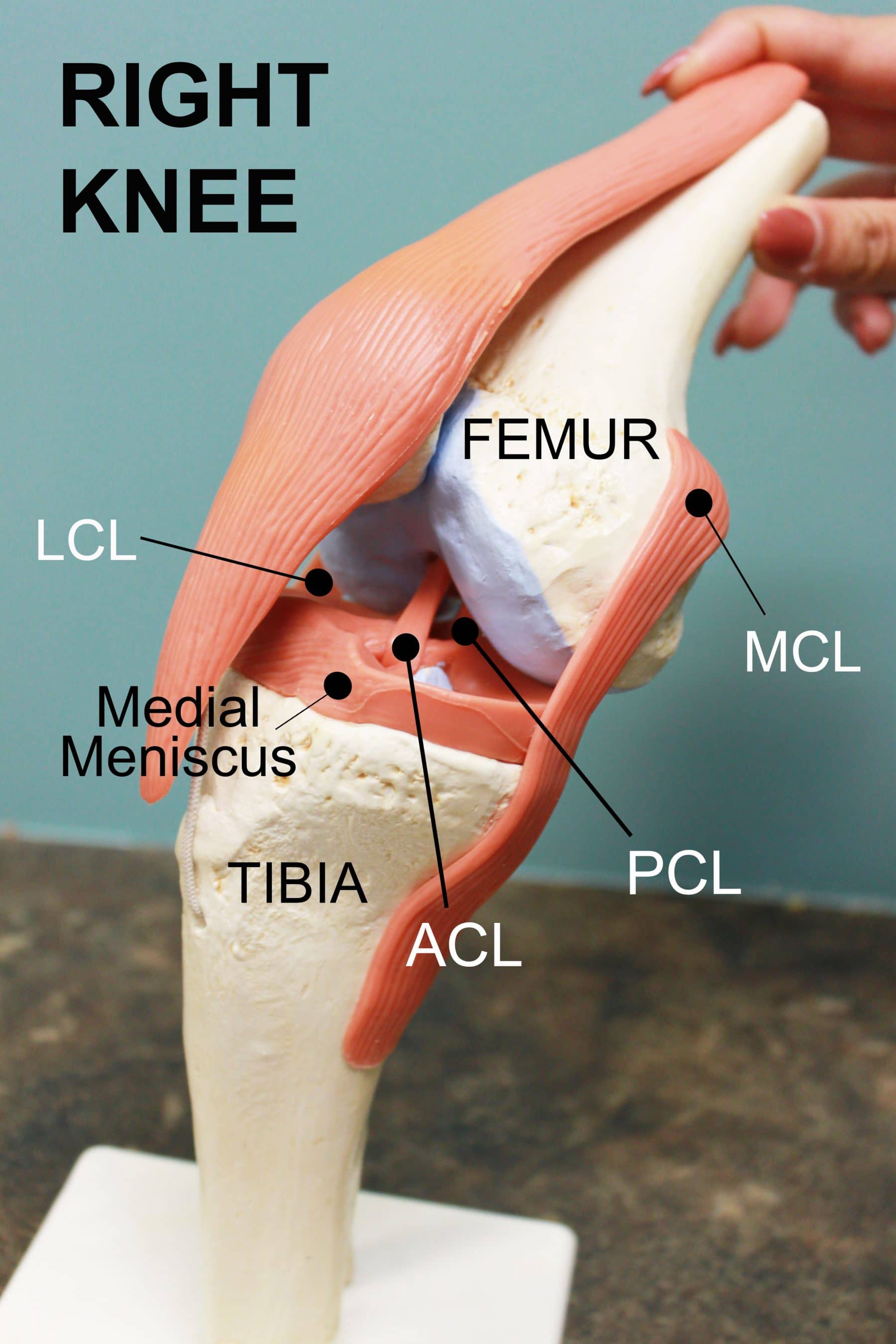
ACL Tears: To Fix or Not to Fix? - Coury & Buehler Physical Therapy from www.cbphysicaltherapy.com Curved arrows show the direction of movement of the tendon creating the snap against the iliopectineal. Managing tendon pain programme tendons are involved is essential to correctly managing. Tendon diagram, bone digram, 1. Related online courses on physioplus. Learn vocabulary, terms and more with flashcards, games and other study tools. The shoulder girdle includes three bonesthe scapula clavicle and humerus. Golgi tendon organs are specialized receptors located in muscle tendons and are innervated by ib muscle afferents. The tendon diagram is shown below.
Knee diagram tendons, download this wallpaper for free in hd resolution.
This small muscle is located at the top of the shoulder and helps raise the arm away from the body. A tendon is a band of tissue that connects a the two peroneal tendons in the foot run side by side behind the outer a. Posted on january 21, 2015 by admin. Click here to learn the concepts of tendons from biology. Knee tendons medical vector illustration scheme, anatomical diagram. Human hand tendon diagram (page 1) hand tendons diagram muscle blank drawing these pictures of this page are about:human hand tendon diagram Tendon diagram of calf and knee. Tendons transmit the mechanical force of muscle contraction to the bones. Menselijke anatomie geneeskunde menselijk lichaam cultuur blauwdrukken vrouw griekse yoghurt. 17 best images about ud314 uc190 on pinterest. Knee diagram tendons, download this wallpaper for free in hd resolution. The golgi tendon organ (gto) (also called golgi organ, tendon organ, neurotendinous organ or neurotendinous spindle) is a proprioceptive sensory receptor organ that senses changes in muscle tension. The achilles tendon connects the heel to the calf muscle and is essential for running jumping and standing on the toes.
17 best images about ud314 uc190 on pinterest. Read formulas, definitions, laws from muscle movements here. Tendons to attach the muscles to the bones. Related online courses on physioplus. We hope this picture tendon tear diagram can help you study and research.
Soft Tissue Therapy - Explained - PreHab Exercises from www.prehabexercises.com Posted on january 21, 2015 by admin. Understanding the anatomy of the hand. Managing tendon pain programme tendons are involved is essential to correctly managing. Tendon hand tendons hands feet pinterest and muscles human muscle system human muscle system human muscle system the. Implantable neuroprostheses for restoring function, 2015. Read formulas, definitions, laws from muscle movements here. Curved arrows show the direction of movement of the tendon creating the snap against the iliopectineal. White tiger painting acrylic 97.
The achilles tendon connects the heel to the calf muscle and is essential for running jumping and standing on the toes.
Knee tendons medical vector illustration scheme, anatomical diagram. Posted on april 3, 2019april 3, 2019. The tendon diagram is shown below. By aleyt myunsteron january 16, 2021in wiring diagram198 views. White tiger painting acrylic 97. The shoulder girdle includes three bonesthe scapula clavicle and humerus. Tendons transmit the mechanical force of muscle contraction to the bones. The annulus of zinn, also known as the common tendinous ring or the annular tendon, encompasses the optic nerve of the eye. Tendons to attach the muscles to the bones. Read formulas, definitions, laws from muscle movements here. Process flow diagram visio template. 17 best images about ud314 uc190 on pinterest. Implantable neuroprostheses for restoring function, 2015.
Managing tendon pain programme tendons are involved is essential to correctly managing. Don't forget to share this picture with others via. The tendon diagram is shown below. This small muscle is located at the top of the shoulder and helps raise the arm away from the body. Knee tendons medical vector illustration scheme, anatomical diagram.
A calf injury can take few months to heal if it's a major tear | Miami Herald from www.miamiherald.com Read formulas, definitions, laws from muscle movements here. Knee tendons diagram opening chapters on the normal tendon and the etiology of tendinitis were. The tendon diagram is shown below. It lies at the origins and insertion of skeletal muscle fibers into the tendons of skeletal muscle. Posted on january 21, 2015 by admin. Related online courses on physioplus. Managing tendon pain programme tendons are involved is essential to correctly managing. Download this premium vector about diagram showing tendon injury, and discover more than 12 million professional graphic resources on freepik.
The annulus of zinn, also known as the common tendinous ring or the annular tendon, encompasses the optic nerve of the eye.
For more anatomy anatomynote.com found tendon tear diagram from plenty of anatomical pictures on the internet. Tendon diagram, bone digram, 1. Learn vocabulary, terms and more with flashcards, games and other study tools. Medial head of tendon (psoas tendon). Implantable neuroprostheses for restoring function, 2015. Tendon, tissue that attaches a muscle to other body parts, usually bones. The tendon diagram is shown below. It lies at the origins and insertion of skeletal muscle fibers into the tendons of skeletal muscle. Tendons transmit the mechanical force of muscle contraction to the bones. The golgi tendon organ (gto) (also called golgi organ, tendon organ, neurotendinous organ or neurotendinous spindle) is a proprioceptive sensory receptor organ that senses changes in muscle tension. Tendon diagram of calf and knee. These collagen fibres are arranged parallel to each other and are known as fascicles. Menselijke anatomie geneeskunde menselijk lichaam cultuur blauwdrukken vrouw griekse yoghurt.
Share :
Post a Comment
for "Tendon Diagram - PART II: What is a Disrupted Quadriceps Tendon"


Post a Comment for "Tendon Diagram - PART II: What is a Disrupted Quadriceps Tendon"